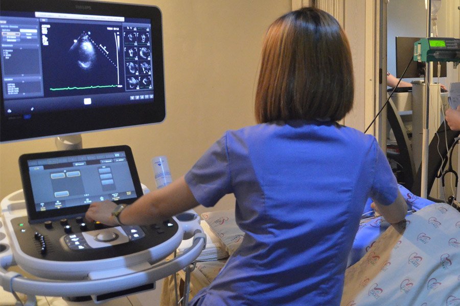
2D Echo Service
2D Echocardiography (2D Echo) is a non-invasive imaging technique used to visualize the heart's structure and function in real-time. It employs high-frequency sound waves (ultrasound) to create two-dimensional images of the heart, allowing healthcare providers to assess various cardiac conditions.
Key Features of 2D Echocardiography :
Purpose:
- Evaluate cardiac structure: To assess the size, shape, and thickness of the heart chambers, valves, and surrounding structures.
- Assess heart function: To evaluate the heart's pumping ability, including systolic and diastolic function.
- Detect abnormalities: To identify congenital heart defects, valve disorders, cardiomyopathies, and other structural heart diseases.
- Monitor heart conditions: To follow up on known cardiac issues and assess the effectiveness of treatments.
Indications for 2D Echo :
- Symptoms of heart disease: Such as chest pain, shortness of breath, palpitations, or unexplained fatigue.
- Heart murmurs: Detected during a physical examination to evaluate potential valvular heart disease.
- Congestive heart failure: To assess the heart's function and determine the cause of heart failure.
- Preoperative assessment: To evaluate cardiac status before surgery.
- Follow-up on previous echocardiograms: To monitor changes in heart structure or function over time.
Procedure:
- Patient preparation: The patient typically lies in a left lateral decubitus position (on their left side) with the chest exposed. No special preparation is generally required.
- Ultrasound technique: A gel is applied to the chest wall to facilitate sound wave transmission. A handheld transducer is moved over the chest to obtain images of the heart from different angles.
- Image acquisition: Multiple views of the heart are captured, including the parasternal, apical, and subcostal views.
What is Assessed :
- Heart chambers: Evaluating the size and function of the left and right atria and ventricles.
- Valves: Assessing the structure and function of the heart valves (mitral, aortic, tricuspid, and pulmonary) for abnormalities such as stenosis or regurgitation.
- Wall motion: Observing the movement of the heart walls to identify areas of decreased motion or ischemia.
- Ejection fraction: Calculating the percentage of blood ejected from the ventricles with each heartbeat to assess overall heart function.
Common Conditions Detected:
- Valvular heart disease: Including aortic stenosis, mitral regurgitation, and other valve abnormalities.
- Congestive heart failure: Identifying causes and assessing the severity of heart failure.
- Cardiomyopathy: Evaluating different types of cardiomyopathy (e.g., hypertrophic, dilated).
- Congenital heart defects: Detecting structural heart defects present at birth.
- Pericardial effusion: Identifying fluid accumulation around the heart.